The findings on the effects of glutathione levels on cell signaling pathways provide new ways to intervene in the aging process and treat degenerative diseases.
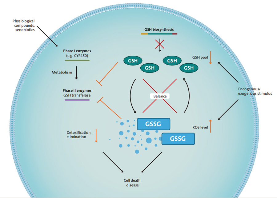
Oxidative stress, an imbalance between the production of reactive oxygen species and the cells’ antioxidant defenses, associated with human disease and aging.
Oxidative stress can lead to apoptosis or cell death. Acute degenerative diseases, such as stroke, arteriosclerosis, diabetes, Alzheimer’s disease, and Parkinson’s disease, can develop as a result.
We offer assay systems for the detection of GSH, mitochondrial function, ROS changes, and the detection of REDOX glutathione ratio, which can used as an indicator of cell health.
Oxidative stress analysis
Glutathione is the most important and powerful antioxidant in the cell.
Glutathione is also involved in the second stage of biotransformation.
It can appear in reduced form as a monomer (GSH) or in oxidized form as a dimer (GSSG).
The ratio of reduced GSH to oxidized GSSG is an indicator of oxidative stress. Glutathione supplier
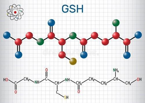
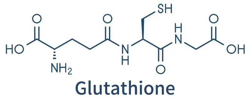
Glutathione composed of three amino acids, glutamic acid, cysteine and glycine. In addition to being the main component of the reduction pool, GSH is probably the most important reserve of the amino acid cysteine.
To prevent oxidative stress caused, for example, by reactive oxygen species (ROS), glutathione oxidized and converted from its reduced monomer form to its oxidized dimer form, GSSG.
Glutathione reductase regenerates two molecules of GSH from GSSG, consuming energy in the process.
98% of GSH in the body is in reduced form.
Glutathione level detection
The change of GSH level is very important for the evaluation of cytotoxic response, and is an important indicator of oxidative stress, which may cause apoptosis or cell death.
The kit provides a simple and rapid microplate detection scheme to directly detect the level of total glutathione (GSH+GSSG) and oxidized glutathione (GSSG) in microplate cultured cells, and the ratio of GSH or GSHIGSSG. The results obtained can not only reflect the level of cell health or oxidative stress, but also used for drug development and screening. New compounds have been discovered that can affect the level of GSH in cells.
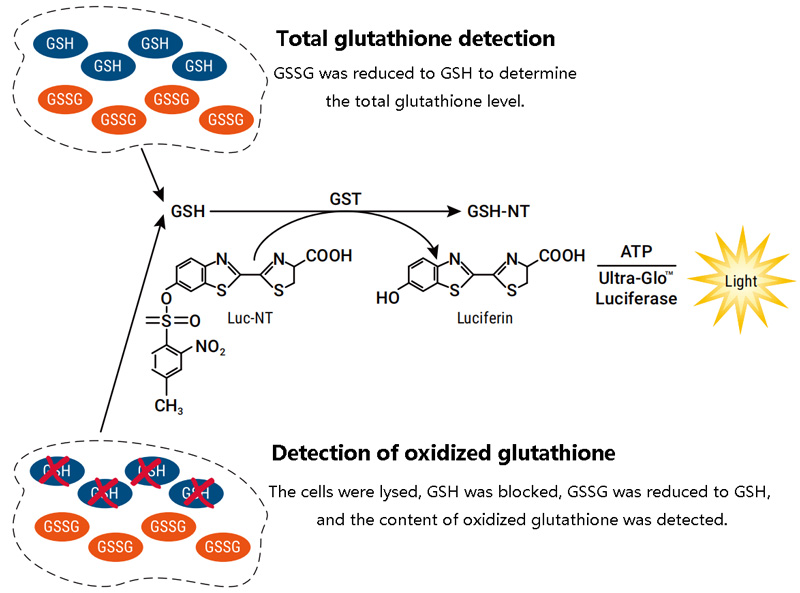
Detection principle
Glutathione detection:
The GSH probe (luciferin) converted to luciferin under the catalysis of GSH S-transferase and coupled with firefly luciferin reaction, and the final detected stable luminescence signal is proportional to the amount of GSH present in the sample.
Glutathione /GSSG detection:
The total glutathione and GSSG detection reactions carried out in parallel, and one reaction used to detect the amount of total glutathione by converting all glutathione (GSH+GSSG) in the cell lysate to reduced glutathione GSH with a reducing agent.
In another reaction, GSSG reduced to GSH by blocking all GSH with a reagent while leaving GSSG unchanged,
and the amount of oxidized glutathione GSSG was quantified by a luminescent reaction.
Since these assays performed directly in a culture hole containing cells,
the loss of glutathione or GSSG minimized and variability reduced.
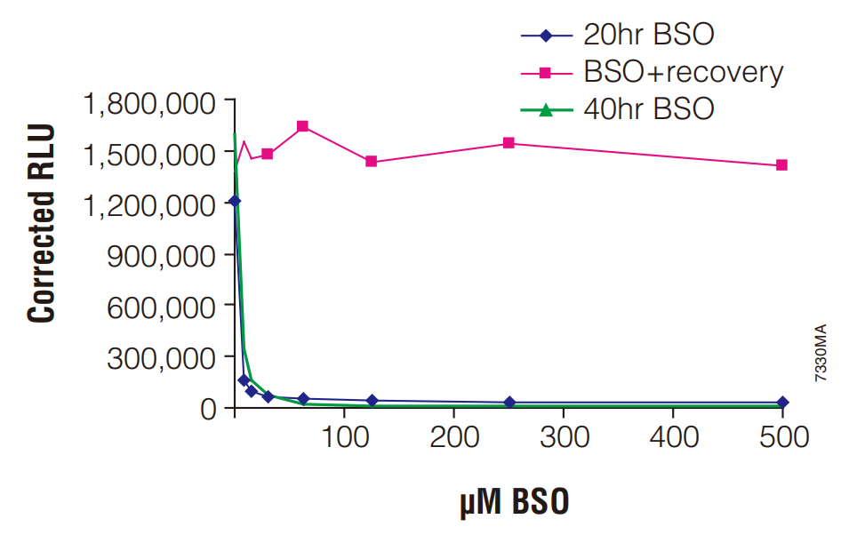
Application: To detect the change of cellular glutathione induced by the compound
The sensitizing compound sulfoxylamine butylthiamine BSO inhibits the depletion and recovery of GSH in cells
Hela cells (5000 cells/well on a 96-well plate) treated with BSO,
which inhibited GSH synthesis, thereby reducing the cell GSH level.
20hr BSO: GSH detected after cells were treated with BSO for 20 hours.
BSO +Recovery: Cells treated with BSO for 20 hours, then washed twice with PBS and fresh BSO-free medium added. GSH in the cells detected after 40 hours.
40hr BSO: Cells treated with BSO, washed after 20 hours, and then added to fresh media containing BSO. GSH in the cells was detected after 40 hours.
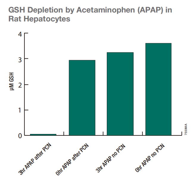
Acetaminophen (APAP) consumes GSH in liver cells
Glutathione Assay used to determine the GSH level in the lysate of adherent cells in 24-well plates.
The GSH standard curve obtained by the detection system used to determine the GSH concentration by interpolation.
GSH decreased significantly after 2 days of treatment with P450 inducer 30μMpregnenolone 16a-carbonitrile (PCN) followed by 5mM paracetaminophen (APAP) for 3 h. However, there little change in GSH level caused by APAP or PCN treatment alone.
Effects of menadione on oxidative stress in A549 lung cancer cells – GSH/GSSG ratio detection
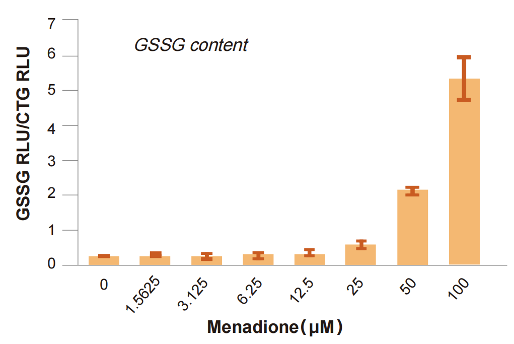
Above: 5000 cells/Wells treated with gradiently-diluted menadione (60 min, 37 ° C). After GSSG detected, the data normalized with the viability test values using a reagent (CTG) to compensate for the toxic effects due to menadione and experimental fluctuations.
Menadione had toxic effects at the two highest concentrations shown after 60 minutes.
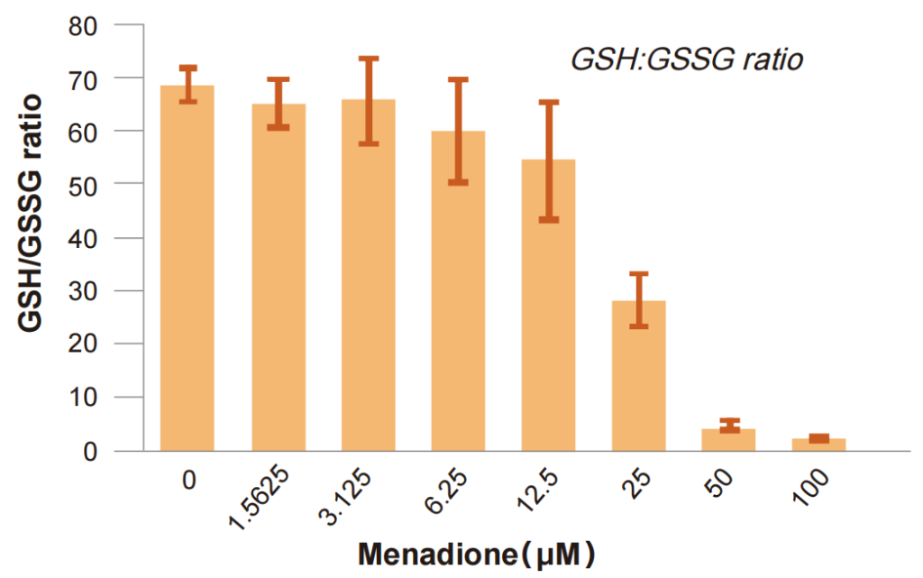
Above: The GSH/GSSG ratio determined by measuring GSSG and total glutathione in A549 cells. Menadione had a significant effect on the REDOX state of cells at higher concentrations.